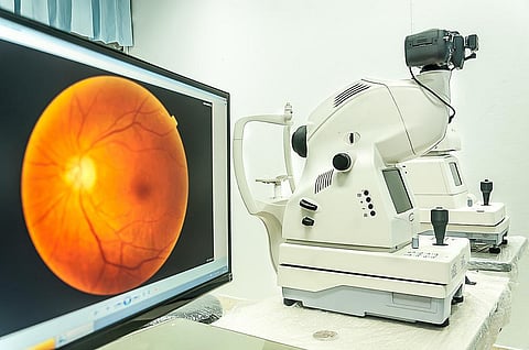

TUESDAY, June 23, 2026 (HealthDay News) -- Colored fundus photography (CFP) can identify regions of the retina associated with risk factors for Alzheimer disease (AD), according to a study published online June 16 in the Journal of Alzheimer's Disease.
Seowung Leem, Ph.D., from the University of Florida in Gainesville, and colleagues examined whether deep learning (DL) models can predict AD-related risk factors from CFP and characterized the retinal structures underlying these predictions. DL models were trained to predict 12 factors linked to AD pathology or incidence using U.K. Biobank CFPs: six categorical and six continuous factors. Model performance, model saliency, and saliency-derived scores were assessed and compared to retinal morphometry.
The researchers found that the predictive performance of DL varied from an area under the receiver operating curve of 0.5654 to 0.9480 and R2 from −0.0291 to 0.7620 for categorical and continuous factors, respectively, outperforming most of the morphometry-based machine learning models. Biologically meaningful regions were consistently highlighted with the saliency-based score, especially the optic nerve head and retinal vasculature. There was also alignment for the saliency-based score with present morphometric variations. Significant differences were seen for several saliency-based scores between incident AD and matched controls.
"With the assistance of artificial intelligence, we are now able to identify subtle retinal variations that were formerly overlooked across thousands of subjects, which may function as reliable indicators of future disease risk," Leem said in a statement.
